O Conselho Regional de Enfermagem de São Paulo no ano de 2025, iniciou o projeto Avança + com a finalidade de capacitar os enfermeiros do estado de São Paulo na avaliação vesical para detecção de retenção urinária e confirmação do posicionamento do cateter vesical de demora.
Este projeto esta sendo coordenado pelo líder do Centro de Pesquisa Interprofissional em POCUS (Prof. Dr. Vinicius Batista Santos), que também atua como Conselheiro do Coren-SP e conta com a colaboração de instrutores, pertencentes ao CePI-POCUS.
INFORMAÇÕES SOBRE O PROJETO AVANÇA +
O Conselho Regional de Enfermagem de São Paulo (Coren-SP) apresenta o subprojeto AVANÇA+ na Ultrassonografia Vesical, uma ação de educação permanente que capacita enfermeiros para o uso da ultrassonografia à beira leito na avaliação do volume vesical. A prática contribui para a prevenção de infecções do trato urinário (ITU) ao reduzir o cateterismo vesical desnecessário e apoia a identificação de diagnósticos de enfermagem, como Eliminação urinária prejudicada, promovendo um cuidado mais seguro e baseado em evidências.
O curso é voltado a enfermeiros de todos os níveis assistenciais — hospitalar, pré-hospitalar, atenção básica e domiciliar — e busca fortalecer o protagonismo da enfermagem na vigilância de riscos e na tomada de decisão clínica fundamentada em dados objetivos.
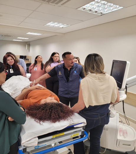
O projeto tem como objetivo capacitar os enfermeiros para a realização da ultrassonografia vesical à beira leito, desenvolvendo o raciocínio clínico e a autonomia profissional frente aos achados da avaliação. Entre os objetivos específicos estão: habilitar o profissional na identificação de diagnósticos de enfermagem relacionados à eliminação urinária, incentivar a adoção de protocolos de prevenção de infecção e estimular a incorporação da ultrassonografia vesical como prática rotineira.
A formação possui 8 horas presenciais, divididas em 4 horas teóricas, com conteúdos sobre anatomia, fisiologia e prevenção de IRAS, e 4 horas práticas, com simulações e manuseio supervisionado dos equipamentos. A modalidade é híbrida, com parte teórica online e prática nas dependências do Coren-SP ou em unidades parceiras.
Os participantes com inscrição ativa no Coren-SP serão avaliados por prova teórica e checklist prático. Os aprovados receberão certificação oficial do Coren-SP, válida como título de educação continuada, reforçando a valorização profissional e o compromisso com a qualidade e segurança do cuidado.
Para mais informações acesse o site do COREN-SP
I